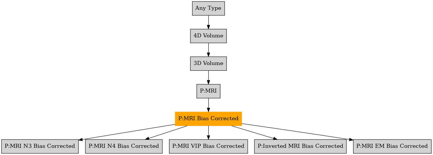

Defined in file : brainvisa/toolboxes/primatologist/types/primatologist.py
Cortex Topological Correction
Hemi Surfaces and Sulci Extraction
Histogram Analysis (for Morphologist)
Invert
Pial Mesh
Prepare For Morphologist
Skull-Strip MRI
Sulci Skeleton
Surfaces and Sulci Extraction
{Viewer} Bias Corrected MRI
{Viewer} Brain Mask
{Viewer} Cortex Roots
{Viewer} Cortex Skeleton
{Viewer} Grey White Mask
{Viewer} Mid Interface
{Viewer} Parcellation
{Viewer} Probabilities
{Viewer} Registered Atlas Labels
{Viewer} Registered Atlas Lateralization
{Viewer} Registered Atlas Template
{Viewer} Skull Stripping Mask
{Viewer} Split Brain Mask
{Viewer} Sulci Voronoi
{Viewer} Topology
Aperio svs
BMP image
BrainVISA volume formats
DICOM image
Directory
ECAT i image
ECAT v image
FDF image
FreesurferMGH
FreesurferMGZ
GIF image
GIS image
gz compressed MINC image
gz compressed NIFTI-1 image
Hamamatsu ndpi
Hamamatsu vms
Hamamatsu vmu
JPEG image
Leica scn
MINC image
NIFTI-1 image
PBM image
PGM image
PNG image
PPM image
Sakura svslide
SPM image
TIFF image
TIFF(.tif) image
Ventana bif
VIDA image
XBM image
XPM image
Zeiss czi
{center}/{subject}/anatomical_mri/{acquisition}/pre_analysis/nobias_<subject>
{center}/{subject}/anatomical_mri/{acquisition}/{analysis}/nobias_<subject>
time_point , time_duration , rescan , mapped , acquisition_date , contrast , center , subject , acquisition , analysis